
Tympanoplasty -various aspects-
The tympanic membrane (TM) may become damaged and become perforated, which will not heal, as a result of chronic suppurative otitis media (CSOM), which is characterised by recurrent or protracted acute otitis media episodes lasting longer than 12 weeks. Tympanoplasty is a surgical treatment used to replace the ossicles after a ruptured tympanic membrane has been repaired in order to prevent reinfection and restore hearing. Tympanoplasty uses endoscopic and microscopic techniques. The interprofessional team’s role in diagnosing and treating tympanic membrane perforations is highlighted in this activity, which discusses the reasons for tympanoplasty and available treatments.
Before going through this article let us go through the common questions arising in the minds of people regarding Tympanoplasty surgery .
What is a tympanoplasty surgery?
The surgical treatment known as tympanoplasty is used to repair a perforated tympanic membrane, either with or without ossicle rebuilding, in an effort to stop reinfection and restore hearing.
What are the five types of tympanoplasty?
The Wullstein Classification
Type I: just the TM is corrected; there are no middle ear abnormalities.
Type II includes middle ear repair and TM; the malleus is debilitated.
Type III: the TM is fixed to the stapes head, but the malleus and incus are deformed.
Type IV: The movable stapes footplate is grafted with the TM.
Does tympanoplasty improve hearing?
When a hole in the eardrum (also known as the tympanic membrane) doesn’t close on its own, ENT surgeon performs a tympanoplasty. It is done to repair the deficit in the continuity of tympanic membrane and to enhance hearing.
Is tympanoplasty surgery painful?
It’s normal to experience mild, sporadic ear ache in the first two weeks following surgery. The pain above or in front of the ear rises when chewing and can be either transient or stabbing in character. You may take the painkiller your ENT surgeon prescribes for a few days following surgery.
Is tympanoplasty a major surgery?
As with any operation, there are risks associated with tympanoplasty, including bleeding and infection. Facial nerve palsy can occur if due care is not taken by the operating ENT surgeon . On the other hand, problems following tympanoplasty surgery are quite uncommon. Vertigo are some other potential issues. Vertigo can occur if the footplate is damaged by the operating ENT Surgeon-which is very rare .Giddiness due to anesthesia side effects can occur which subsides in 12 hrs .
Is tympanoplasty expensive?
=The price range for a Tympanoplasty Eardrum Repair in India is between Rs 25000 -Rs 45000 .If any patient needs to get operated for Tympanoplasty surgery ,he may take appointment from ENT specialist doctor Dr Sagar Rajkuwar at the following adress-
Prabha ENT clinic, plot no 345,Saigram colony, opposite Indoline furniture Ambad link road ,Ambad ,1 km from Pathardi phata Nashik -Dr Sagar Rajkuwar (MS-ENT), Cel no- 7387590194,9892596635
Clinic website -www.entspecialistinnashik.com
How many hours is tympanoplasty surgery?
Typically, a patch myringoplasty takes 15-20 minutes. For children, tympanoplasty can take one to two hours. Following surgery, you will have a conversation with your doctor. Following surgery, your kid will awaken in the recovery area.
Can I walk after tympanoplasty?
To begin, walk a little bit more than you did the previous day. Gradually raise the distance you walk. In addition to increasing blood flow, walking guards against constipation and pneumonia. For the first two or three days following surgery, avoid leaning over and making abrupt head movements.
What are the side effects of tympanoplasty?
Complications and Side Effects of Tympanoplasty
vomiting and nausea throughout the first 48 to 72 hours.
For a period of three to five days: mild to moderate discomfort at the site of the incision or in the ear. A mild case of fever. feeling lightheaded or unsteady for a few days ,reduced hearing in the ear that was operated on for a few weeks.
What is the success rate of tympanoplasty?
The literature reports a good success rate for the closure of simple tympanic membrane perforations [4,5, 6]. Success rates range from 35 to 94% for children and from 60 to 99% for adults, according to several studies [6, 7]. Different writers define success in tympanoplasty differently.
What age is tympanoplasty for?
After the age of four, tympanoplasty is performed according to their clinical algorithm. ENT Surgeon will treat the nose ( Adenoidectomy and Tonsillectomy is done if the patient is symptomatic and changes are seen in CT SCAN-PNS and then only Tympanoplasty is done .It is done only when the ear is dry .
What happens if tympanoplasty fails?
The greatest danger associated with tympanoplasty is failure; in the event that the surgery is unsuccessful, the ear hole will remain as it was previously
What not to do after tympanoplasty?
=For the first two or three days following surgery, avoid leaning over and making abrupt head movements. These movements could cause vertigo. For approximately two to four weeks, or until your doctor gives the all clear, refrain from doing any intense activity, such as jogging, weight lifting, riding a bicycle, or aerobic exercise.
Can I sleep on my side after tympanoplasty Right After Surgery ?
For the first week following surgery, you should rest with your head propped up in a chair or with two pillows at minimum. For around two weeks, try sleeping with your head resting on the pillow rather than your face.
| ReplyForward |
Introduction
The tympanic membrane (TM) may become damaged and become perforated, which will not heal, as a result of chronic suppurative otitis media (CSOM), which is characterised by recurrent or protracted acute otitis media episodes lasting longer than 12 weeks.The most prevalent infectious disease affecting children worldwide is CSOM. Upper respiratory infections, poor nutrition, unhygienic living conditions, family history, low birth weight, cranial deformities, and having Native American, Native Alaskan, or Aboriginal Australian ancestry are all risk factors. Otorrhoea that enters the external ear canal via the TM perforation and hearing loss are signs of CSOM. Even while the hearing loss is normally quite minor (10 to 20 dB), it could go worse with extensive perforations. In other instances, ossicular chain degradation can also happen, which can have a more significant impact on the audiology. (50 to 70dB). In such cases, it is vital to rule out the occurrence of cholesteatoma. Both the pars tensa and the pars flaccida have the potential to have tympanic membrane perforations. (though the former is by far the most common). Depending on where it is in relation to the annulus, the hole can also be classified as marginal or central and wet or dry. (persistent otorrhoea or no active otorrhoea, respectively).
CSOM with cholesteatoma is a subtype of the condition. Cholesteotoma requires mastoid surgery with tympanoplasty .Middle ear cholesteatomas are most frequently a result of an acquired disease condition. They are made up of a sac of squamous epithelium that often develops in the attic and originates at the pars flaccida of the TM. These sacs can extend outside of the TM, eroding bone and leading to the breakdown of ossicular chains. Cholesteatoma can present with the same signs and symptoms as CSOM, such as foul-smelling otorrhoea, hearing loss, TM perforation, and attic retraction.
With or without reconstructing the ossicles (ossiculoplasty), a perforated TM is surgically repaired with a tympanoplasty in an effort to stop reinfection and restore hearing. The most frequent indication is CSOM; however, massive invasive cholesteatomas may necessitate mastoidectomy and TM reconstruction. The first tympanoplasty procedures were performed in the 1950s by Wullstein and Zollner, who popularised the use of overlay grafts to repair perforated TMs and restore the middle ear’s sound-conduction system. Tympanoplasty surgical techniques have since undergone modifications, as discussed in this article.
Classification by Wullstein-
The Wullstein classification of tympanoplasty divides it into five categories.
Type I: TM repair alone; no abnormalities in the middle ear. Myringoplasty and type I tympanoplasty are interchangeable terms.
Type II: the malleus is eroded; the TM and middle ear are repaired. To perform a tympanoplasty, the TM is grafted to the incus.
Type III: the malleus and incus have a fault, and the TM is repaired onto the stapes head.
Type IV: the TM is attached to the movable stapes footplate.
Type V: the stapes footplate is fixed during the repair.
Biology and Anatomy
The petrous temporal bone contains the middle ear cleft, sometimes referred to as the tympanic cavity. The inner layer of the TM, the Eustachian tube opening, and the ossicular chain are the anatomical parts of the middle ear. (malleus, incus, stapes). The Eustachian tube connects anteriorly with the nasopharynx, allowing the middle ear to equalise pressure with the surrounding environment. The middle ear cleft communicates posteriorly with the mastoid air cells via the tympanic antrum and the aditus ad antrum.
Before understanding Tympanoplasty surgery it is important to know first about the anatomy of the middle ear. The majority of the lateral wall of the middle ear fissure is formed by the TM. The fibrous stratum (lamina propria) middle layer, which is the most significant component and gives the TM stability, the stratified squamous epithelial outer layer that is continuous with the external ear canal, and an inner layer that is continuous with the cuboidal mucosa of the middle ear make up this structure. Solid collagen fibres are present in the fibrous stratum, and it is their restricted capacity for stretching that allows for high compliance with very small acoustic pressure displacements but resistance (low compliance) to further stretching at greater pressures. The TM has a diameter of around 10 mm, a thickness of about 0.1 mm, and is typically pearly grey and just slightly translucent, allowing for the visibility of ossicular structures.
Anatomically, the TM is divided into the superiorly located pars flaccida and the inferiorly located pars tensa. The pars flaccida lies anterior and posterior to the malleolar ligaments; it is thinner and more flexible than the pars tensa because it lacks a core fibrous layer. The majority of the TM is made up of the pars tensa, which covers the region beneath the malleus’ neck. In clinical practice, the TM is further divided into four quadrants by two fictitious lines that pass through the umbo horizontally and the malleus handle perpendicularly, respectively. Understanding the intricate spatial relationships between the middle ear’s parts is crucial for otologic surgeons, and the middle ear’s functional and anatomical qualities frequently impact surgery.
The middle ear apparatus’s vibrational driver in terms of function is the TM. A “liquid wave” is created when sound energy, in the form of air pressure waves, is transported from the TM through the ossicular chain to the fluid-filled inner ear via the oval window. The TM and ossicular chain’s function is to amplify sound energy and send this magnified signal to the cochlea, which uses specialised cells (hair cells) arranged tonotopically along the basilar membrane to transform this mechanical energy into electrical nerve impulses. The cochlear nerve then relays these signals to the brainstem.
Indications-
Tympanoplasty aims to repair the TM with or without repairing the ossicular chain, preventing recurrent infections (otorrhoea), and restoring hearing in the process. The main symptoms are CSOM or CSOM along with cholesteatoma. A documented cause of tympanoplasty in the literature is iatrogenic perforation in paediatric patients undergoing ventilation tube insertion for otitis media with effusion. Repair may also be necessary for traumatic TM perforations caused by mechanical damage that do not heal (acute perforations normally heal on their own in 80% of patients).
Contraindications
Tympanoplasty contraindications can be classified as either absolute or relative. Patient age (at the extremes of age), a non-functioning Eustachian tube, a dead (non-hearing) ear, or consideration of tympanoplasty in the patient’s better hearing ear are relative contraindications. Uncontrolled cholesteatoma, uncontrolled otitis media complications or sequelae, such as meningitis or lateral sinus thrombosis, cancer, or patient-specific anaesthetic contraindications are examples of absolute contraindications for tympanoplasty .
Equipment for tympanoplasty surgery-
The following tools can be required to complete a tympanoplasty:
Zero degree endoscope
The following may also be necessary if the tympanoplasty is done endoscopically:
Camera with high resolution, light source, and monitor
Unflexible endoscope (0, 30, and 45 degrees)
Personnel-
A tympanoplasty requires the following personnel:
the otologist
an anaesthetist.
Scrub team planning
A number of tests are done to evaluate the middle ear’s functionality before to surgery, and the results can have an impact on both the tympanoplasty technique employed and the postoperative result.
Any patient who presents with otorrhoea and hearing loss should first have a thorough medical history taken, followed by a focused head and neck examination and an otoscope/microscope examination of the ears. This will provide crucial details on the TM perforation’s size, location (which was previously classified into four quadrants), the type of otorrhoea (wet or dry), and whether or not there is now an active inflammatory response. Tympanography, pure tone audiometry (PTA) with speech discrimination, and tuning fork evaluation (Rinne and Weber tests at 512 Hz) should be carried out both before and after surgery to compare the differences between air and bone conduction thresholds. (air-bone gap).
Simple tympanic membrane perforations seldom require imaging, but if they do, computed tomography (CT) is the ideal imaging technique. Planning the surgical therapy of cholesteatoma typically involves using CT scanning of the temporal bone.
Patients receiving tympanoplasty are increasingly using questionnaires to assess their health-related quality of life in addition to clinically evaluating their middle ear. The majority of validated surveys assess four dimensions: impact on daily life and interpersonal connections, functional disability, psychological health, and physiological complaints. Currently, they are not a required component of the tympanoplasty preoperative workup; nevertheless, future advancements in psychometric measurement techniques are anticipated to produce results that are both scientifically sound and trustworthy.
Patients will give their permission for tympanoplasty prior to surgery. Risks include discomfort, bleeding, infection, graft failure, recurrence, further surgery, severe hearing loss or deafness, disorientation, and injuries to the chorda tympani nerve or the facial nerve leading to facial palsy. Patients will have the chance to talk with an anaesthetist before surgery, and the treatment is normally carried out under general anaesthesia.
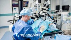
Technique for tympanoplasty surgery–
A postauricular approach is traditionally used for the bulk of middle ear surgery involving the TM; endaural (via the ear canal) and transcanal procedures are also employed. Which procedure to utilise depends on a number of parameters, including the size of the TM hole, the size of the ear canal, and the surgeon’s preference. Each technique has benefits as well as drawbacks. Trans-canal or endoscopic ear surgery is becoming more and more common thanks to a trend towards less intrusive surgical methods, similar to that seen in other surgical specialties.
Regardless of the tympanoplasty procedure employed, standard theatre preparations are carried out. It is normal practise to provide prophylactic antibiotics. (although less so during endoscopic surgery). A head ring is used to give support whilst the patient is lying supine on the operating table. If necessary, the patient will have their postauricular hair shaved. Local hemostasis must be accomplished before administering local infiltration (lidocaine with adrenaline).Prior to starting the treatment, the ear canal is inspected, cleansed as necessary, and the TM perforation and ossicles are examined under microscopic or endoscopic vision.
Grafts for tympanoplasty surgery–
Since autologous material for grafting is widely accessible, affordable, and biocompatible, there is little need for using artificial substitutes. However, studies have also documented employing fascia lata, canal skin, and periosteum. Autologous grafts are typically generated from temporal fascia or tragal/conchal perichondrium. The main benefits of employing alloplastic grafts, such as acellular dermal matrix and absorbable gelatin sponge, include the lower morbidity of autologous graft harvesting, which includes less discomfort and better cosmetic results without further scars. The above-mentioned benefits could be offset by the expense and risk of infectious disease transmission associated with employing these grafts for tympanoplasty surgery . Many otologists prefer to employ autologous grafts in actual practice.
The temporalis fascia is more frequently employed in tympanoplasty surgery than cartilage, and its success rates for tympanoplasty range from 93% to 97%. However, cartilage and perichondrium can be collected by making an incision through the medial side of the tragal skin, perichondrium, and cartilage in circumstances calling for higher stability, such as multiply-recurrent perforations or high-risk patients. While it has been discovered that the use of cartilage is more successful than temporal fascia, primarily because of its high tensile strength, which makes it more resistant to shrinkage, on occasion questions have been raised regarding sound conduction properties due to its rigid quality (resulting in a potential mild, low-frequency, conductive hearing loss from a mass effect) and potential to be mistaken for cholesteatoma postoperatively.
Microscopic Strategy for tympanoplasty–
The postauricular and trans-canal techniques are widely employed for tympanoplasty while utilising a microscope. Although far less frequently utilised in contemporary tympanoplasty, the Lempert endaural technique is still a viable option. The ear is folded anteriorly, and the surgery begins with the surgeon creating a semicircular incision about 1 cm posterior to the auricle skin fold. A musculoperiosteal flap is created when the incision is carried into the musculoperiosteum and elevated in the direction of the membranous ear canal, where it enters the bony ear canal. Until the surgeon reaches the incisions for the tympanomeatal flap, the skin along the posterior portion of the bone canal is elevated. The middle ear can then be accessed by raising the tympanomeatal flap.
An ossiculoplasty can be done now, if necessary, to restore the ossicular chain. Using cup forceps or scissors, the TM perforation edge is refreshed. The TM is rebuilt using an underlay approach in which the limbus is undermined, the TM is elevated, and the perforation is medially closed with a graft. The autologous graft must stabilise the ear canal and cover the TM defect for the procedure to have the best probability of success. When lifting the limbus from its bone sulcus, caution should be exercised to prevent injuring the chorda tympani in the back area.
Endoscopic Method for tympanoplasty –
TM perforations can be repaired endoscopically, which is less invasive than conventional postauricular and endaural methods. By avoiding external incisions, it can give a more comprehensive picture of the middle ear anatomy and shorten the time required for surgery and recovery.Studies have shown that microscopic tympanoplasty and perforation closure rates for audiologic recovery are comparable. One-handed surgical operations and potential damage to nearby structures from heat produced by the endoscope’s light source are drawbacks of endoscopic middle ear surgery.
A transcanal technique is used when doing endoscopic tympanoplasty. The perforation’s margins are once again de-epithelized as necessary. The tympanomeatal flap and annulus can be raised by means of an ear canal incision (endaural, lateral circumferential, or swing door), which also provides access to the middle ear. Peeling the malleus from the TM occurs at this time, and if necessary, the ossicular chain is repaired (ossiculoplasty). The prepared graft is positioned lateral to the malleus and medially to the TM remnant. The middle and outer ear canals are treated with gel foam sponges. More recently, a “butterfly cartilage tympanoplasty technique” that avoids raising the tympanomeatal flap has been described in the literature. A graft is inserted into the hole after the creation of cartilaginous pseudo-flanges, with one flange medial to the TM and the other lateral. Gel foam sponges are positioned around the graft’s border in tympanoplasty surgery once an appropriate position has been found.
Complications-
Tympanoplasty is successful in more than 93% of patients, according to studies. (in patients undergoing primary tympanoplasty using temporalis fascia graft). However, problems are possible with any procedure. Recurrence (which includes graft failure), conductive hearing loss, TM perforation, and intra- or postoperative ventilation tube insertion are the most common side effects following tympanoplasty.
Recurrence of TM perforation was reported in one large case series that examined over 1000 patients who had cartilage tympanoplasty in 3.6% to 4.2% of patients (patients with cholesteatoma and high-risk perforations, respectively) and in 1.9% to 11% of patients (high-risk perforations with/without patients with cholesteatoma). (patients undergoing TM repair to improve hearing). Patient comorbidities like diabetes, smoking, and immunosuppression are important contributing factors that can affect the postoperative result in addition to the method and graft of choice.
The chorda tympani and facial nerve are two significant neural structures that cross the middle ear and knowledge of this is important to the ENT surgeon before doing tympanoplasty .The facial nerve passes above the oval window into the temporal bone and continues along the back of the tympanic cavity. Fortunately, there is a limited incidence of iatrogenic facial nerve injury because otology surgeons who perform tympanoplasty have significant training. During middle ear surgery, the chorda tympani will always be there, and in cases of cholesteatoma in CSOM, it might need to be sacrificed to achieve complete disease clearance.
Clinical Relevance-
Tympanoplasty has been carried on on CSOM patients since the 1950s with the goal of lowering infection recurrence, increasing hearing, and ultimately enhancing social development and quality of life. Tympanoplasty techniques have changed over time, however many centres still choose to use a postauricular approach with an autologous graft despite the rise in popularity of endoscopic middle ear surgery.
Improving Healthcare Team Results in Tympanoplasty surgery-
An interprofessional team made up of otologists, audiologists, radiologists, anesthetists, and nurses is necessary to manage patients with CSOM. For the best possible patient outcomes, including lower infection rates and better hearing, prompt diagnosis and careful surgical planning are necessary. Coordinating with speech therapists both before and after surgery may also be necessary for paediatric children whose hearing impairment has the potential to have a significant influence on speech and language development. Nurses will help with patient evaluation, organise tasks for the many specialists, aid with surgery, and provide patient counselling. In order for everyone involved in care to have access to the same patient information to inform therapy decisions, all interprofessional team members must keep accurate and up-to-date records. With the fewest adverse events, the interprofessional approach will produce the best patient outcomes.
If any patient needs to undergo Tympanoplasty surgery or mastoidectomy surgery ,then he may contact ENT specialist doctor Dr Sagar Rajkuwar at the following adress-
Prabha ENT clinic, plot no 345,Saigram colony, opposite Indoline furniture Ambad link road ,Ambad ,1 km from Pathardi phata Nashik ,422010 ,Maharashtra-Dr Sagar Rajkuwar (MS-ENT), Cel no- 7387590194 ,9892596635
Clinic website –
www,entspecialisinnashik.com


