

Tinnitus Pulsatile -various aspects-
Tinnitus with pulse -Pulsatile Tinnitus: What Is It? People who suffer from pulsatile tinnitus frequently experience rhythmic whooshing, throbbing, or thumping in one or both ears. The sounds are said to be grating by some patients. Others, however, find the noises to be incapacitating and intense, making it difficult to focus or rest. Different from the more prevalent, constant form of tinnitus is pulsatile tinnitus. Even though pulsatile tinnitus is frequently harmless, it is more likely to have a specific cause and may be the initial symptom of a more serious underlying condition.

Sometimes lpusatile tinnitus goes away by itself. Patients with pulsatile tinnitus symptoms should, however, get a full medical evaluation because it can be brought on by potentially harmful conditions. Fortunately, once the underlying cause is found, pulsatile tinnitus is frequently successfully treated and cured .Pulsatile tinnitus symptoms -Hearing a constant beat or whooshing sound on a regular basis is the most typical symptom of pulsatile tinnitus. The sound or beat frequently matches the patient’s heartbeat.
Both the beat and the music will speed up as their heart rate rises and calm down, respectively .People with pulsatile tinnitus frequently hear it even when they have not exerted themselves, whereas it is typical for people to hear their heartbeat if their heart is pounding vigorously. Because there are fewer outside noises to cover up the beat or sound, pulsatile tinnitus symptoms might also be more obvious at night while you’re lying in bed .The beat or sound may be constant or intermittent.
The symptoms of pulsatile tinnitus can be distracting and noisy, interfering with daily life for many people
Tinnitus Pulsatile -different facets-
Pulsatile tinnitus is defined as tinnitus with a pulse. Patients with pulsatile tinnitus often have rhythmic throbbing, whooshing, or thumping in one or both ears. Some patients claim that the noise is annoying. However, others find the sounds to be overpowering and debilitating, making it hard to concentrate or relax. Pulsatile tinnitus is distinct from the more common, persistent kind. Although pulsatile tinnitus is usually benign, it is more likely to have a particular cause and might be the first sign of a more underlying ailment.
In some cases, pulsatile tinnitus vanishes on its own. However, because pulsatile tinnitus symptoms can be caused by possibly dangerous diseases, patients should receive a comprehensive medical assessment. Fortunately, pulsatile tinnitus can often be effectively treated and cured once the underlying cause is identified. The most typical symptom of pulsatile tinnitus is experiencing a persistent beat or whooshing noise at regular intervals. The rhythm or tone usually aligns with the patient’s heartbeat.
As their heart rate increases and decreases, the tempo and melody will accelerate and slow down, respectively. In contrast to the common experience of hearing one’s heartbeat when one’s heart is beating hard, individuals with pulsatile tinnitus may experience the condition even in the absence of physical activity. Additionally, pulsatile tinnitus symptoms may be more pronounced at night when you’re in bed because there are fewer external noises to mask the rhythm or tone. The rhythm or sound may be continuous or sporadic.
Pulsatile tinnitus symptoms can be loud and bothersome, making it difficult for some people to carry out their everyday activities.
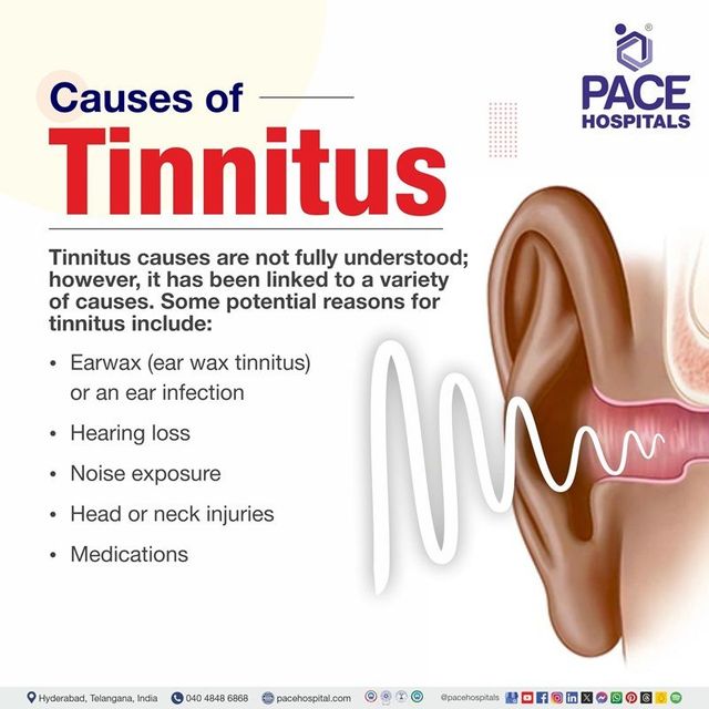
Causes of Pulsating Tinnitus –
Medical experts may frequently pinpoint the underlying health condition that is causing pulsatile tinnitus. Atherosclerosis – Plaques form in the arteries of those who have atherosclerosis. The arteries constrict more when plaque hardens, which restricts blood flow to the body, especially to the head, neck, and ears. The characteristic rhythmic whooshing or thumping sound of pulsatile tinnitus may be produced by one or both of your ears as a result of this. . Blood Vessel Diseases and Malformations – Pulsatile tinnitus can be caused by disorders or abnormalities in the blood arteries and veins, particularly those close to the ears.
Causes of Pulsatile Tinnitus –
Oftentimes, medical professionals are able to identify an underlying health issue that is causing pulsatile tinnitus .Atherosclerosis -People with atherosclerosis develop plaque inside their arteries. Plaque that hardens causes the arteries to become more constricted, which reduces blood flow to the body, particularly to your ears, neck, and head. This could result in one or both of your ears producing the distinctive rhythmic whooshing or thumping sound of pulsatile tinnitus .Disorders and Malformations of the Blood Vessels -Disorders or abnormalities in the blood veins and arteries, particularly those close to the ears, can result in pulsatile tinnitus.
Aneurysms and arteriovenous malformations are just two examples of the anomalies or disorders that can alter the way blood flows through the afflicted blood arteries and can cause pulsatile tinnitus. Various Ear Issues -One of three canals found in the vestibular apparatus of the inner ear is the superior semicircular canal. Pulsatile tinnitus is a common symptom of superior semicircular canal dehiscence syndrome, a disorder in which the portion of the temporal bone that covers the superior semicircular canal is unusually thin or absent. A patient may also hear their heartbeat if the bone covering the major arteries and veins running close to the ear is thinning or missing. Blood pressure is high. Blood flow through the carotid artery is more likely to be turbulent and produce a pulsing sound when blood pressure is high.
Glomus tumours are benign but locally invasive tumours that develop from glomus cells and affect the head and neck. The jugular vein portion located below the middle ear is where these tumours are most frequently found. Glomus tumours can spread to the brain and middle ear. These tumours can produce pulsatile tinnitus and other symptoms when they put pressure on the blood vessels in the head or neck. Due to their strong blood flow, glomus tumours have the potential to result in pulsatile tinnitus just by being near the ear. Intracranial hypertension that is idiopathic .
This is a medical disorder brought on by increased pressure in the cerebrospinal fluid surrounding the brain. Headaches, double vision, soreness behind the eye, and pulsatile tinnitus are some of the symptoms of this increased pressure. Sinus Wall Deviations -Diverticulum and dehiscence of the sigmoid sinus are among these anomalies. A blood vessel on the side of the brain called the sigmoid sinus carries blood from veins inside the brain. The term “sigmoid sinus diverticulum” describes the development of tiny pouches (diverticula) that jut out of the sigmoid sinus wall and into the mastoid bone behind the ear. Dehiscence is the medical term for when the bone that surrounds the sigmoid sinus in the mastoid is missing in part.
Pulsatile tinnitus is the result of these anomalies, which affect the pressure, blood flow, and noise levels within the sigmoid sinus. Additional Roots of Pulsatile Tinnitus -The following diseases can also contribute to the distinctive whooshing or thumping sound of pulsatile tinnitus: -Anemia ,loss of hearing in conductivity, head injury, hyperthyroidism, an enlarged thyroid gland Narrowing of the brain’s blood vessels leaving the brain ,Paget’s illness .
You should have a comprehensive medical evaluation by an otolaryngologist who is knowledgeable about the issue if you believe you have pulsatile tinnitus .About a third of patients may not know what is causing their pulsatile tinnitus, but it is still crucial to rule out any potentially dangerous causes. Additionally, pulsatile tinnitus can be identified using the following imaging techniques: Angiography CTA stands for computerised tomographic angiography.CT scan for computerised tomography ,MRA, or magnetic resonance angiography(MRI) Magnetic resonance imaging a transient bone CT scan ,Ultrasound ,To rule out thyroid or anaemia, blood testing and thyroid function tests may also be required.
Anatomy of inner ear-in relation to pulsatile tinnitus
For update on further important health related topics and frequently asked questions on health topics by general population please click on the link given below to join our WhatsApp group –
https://chat.whatsapp.com/Lv3NbcguOBS5ow6X9DpMMA
Issued in public interest by –
Tinnitus pulsatile pregnancy
Pulsatile tinnitus, characterized by a ringing or whooshing sound in the ears that coincides with the heartbeat, can arise during pregnancy, frequently associated with hormonal changes and increased blood volume, but may also indicate underlying issues such as high blood pressure or preeclampsia.

Here’s a more detailed explanation:
What is Pulsatile Tinnitus?
Pulsatile tinnitus refers to a form of tinnitus (ringing, buzzing, or whooshing in the ears) that synchronizes with the heartbeat.
It may resemble a whooshing or rushing sound, or even mimic a heartbeat within the ear.
Causes of Pulsatile Tinnitus During Pregnancy:
Hormonal Changes:
Pregnancy induces considerable hormonal variations, including heightened progesterone, which can influence blood circulation and possibly result in tinnitus.
Increased Blood Volume:
During pregnancy, the body’s blood volume rises, which can exert additional pressure on blood vessels and potentially trigger pulsatile tinnitus.
High Blood Pressure:
Raised blood pressure, frequently seen during pregnancy, can also play a role in pulsatile tinnitus.
Preeclampsia:
In certain instances, pulsatile tinnitus may indicate preeclampsia, a serious pregnancy complication marked by high blood pressure and organ damage.
Other Conditions:
In rare instances, pulsatile tinnitus during pregnancy could signify other underlying conditions, such as anemia or hyperthyroidism.
What to Do If You Experience Pulsatile Tinnitus During Pregnancy:
Consult with Your Healthcare Provider:
If you notice pulsatile tinnitus during pregnancy, it’s important to notify your doctor or midwife.
Monitor Your Blood Pressure:
Regular monitoring of blood pressure is vital during pregnancy, particularly if tinnitus occurs.
Manage Stress:
Stress can exacerbate tinnitus, so employing relaxation techniques like deep breathing or meditation can be beneficial.
Follow Your Doctor’s Advice:
Your healthcare provider will be capable of evaluating the cause of your tinnitus and suggesting suitable management strategies.
What components make up the inner ear? -In Relation to Tinnitus Pulsatile
Cochlea, labyrinthine canals, and vestibule are the three primary components of your inner ear. Your vestibule and semi-circular canals support your balance, and your cochlea supports your hearing.
What does a cochlea do ? –brief knowledge of this is necessary in relation to pulsatile tinnitus.
Your cochlea is shaped like a snail, tapering from a wide end called the base to a narrow head called the apex. It is a fluid-filled organ. The apex is more sensitive to low-pitched noises, such as a bass drum, whereas the base is most sensitive to high-pitched sounds, such as birds tweeting.
Two slender membranes divide the cochlea into three tubes. The basilar membrane, one of these membranes, resembles an elastic wall, on top of which the organ of Corti is located.
The Corti organ contains minuscule cells known as hair cells -brief knowledge of this is necessary in relation to pulsatile tinnitus. The roughly 18,000 cells in your cochlea are so tiny that they could fit on the head of a pin.
These hair cells are covered in stereocilia- Stereocilia are little, hair-like projections that respond to the movement of the fluid in the cochlea. Inner and outer hair cells are the two different types of hair cells. The outer hair cells respond best to softer noises, while the inner hair cells respond best to louder sounds. Understanding anatomy of inner ear is very important in relation to pulsatile tinnitus .
Although each hair cell has a connection to the hearing nerve, the inner hair cell is primarily in charge of transmitting sound to the brain via the hearing nerve. To quieter sounds, the outer hair cells warn the inner hair cells.
How the cochlea converts sound waves into sounds is as follows-brief knowledge of this is necessary in relation to pulsatile tinnitus.
The small middle ear bones, known as the malleus, incus, and stapes, move as sound travels from your outer ear and strikes your eardrum (also known as the tympanic membrane), which is the wall of your middle ear.
The oval window (a tiny aperture) in the cochlea contains the stapes. Your cochlea’s fluid is rippled when it moves.
Similar to how an ocean current moves plants on the sea floor, this ripple moves the stereocilia.
Your auditory nerve transmits an electrical signal to your brain’s temporal lobe when the stereocilia on your inner and outer hair cells move. The electrical signal is interpreted as sound by the temporal lobe. Tinnitus is related to 8th nerve and pulsatile tinnitus is related to
Semi-circular canals: What are they?-in relation to pulsatile tinnitus.
Your inner ear contains coils of tubes called semi-circular canals. The canals are coated with hair cells and contain liquid, just like the cochlea. These microscopic hairs respond to motions of the body rather than sound waves. They are mostly in charge of rotating motion or motion that is not linear.
DISCLAIMER-Some patients go to net and directly take treatment from there which can lead to catastrophic consequences-Then- Many people ask then why to read all this text -the reason is that it helps you to understand the pathology better ,you can cooperate with treatment better ,your treating physician is already busy with his patients and he does not have sufficient time to explain you all the things right from ABCD ,so it is always better to have some knowledge of the disease /disorder you are suffering from.
What does a vestibule do?- in relation to pulsatile tinnitus.
The vestibule’s utricle and saccule are primarily in charge of up-and-down and forward-and-backward movements.
The vestibular system functions as follows:
- The fluid in your semicircular canals moves the little hairs in the canals when you move your head.
- Your vestibule, which is attached to your semicircular canals via sacs known as your saccule and your utricle, becomes active as a result. Your saccule and utricle, like your semicircular canals, are filled with fluid and have small hairs that enable them to detect movement.
- Your vestibule and semicircular canals communicate the movement to your brain. After that, your brain instructs your body on how to maintain equilibrium.
Issued in public interest by –
Symptoms of pulsatile tinnitus
Pulsatile tinnitus is marked by a rhythmic, pulsing noise in one or both ears that is frequently in time with the heartbeat. It might be characterized as a swooshing, whooshing, or pulsing noise. In some instances, people may experience additional symptoms such as headaches, dizziness, or vision issues, particularly if there is heightened pressure in the skull.
Even in the absence of external noise, tinnitus is typically characterized as a ringing in the ears. However, tinnitus can also produce other kinds of phantom sounds in your ears, such as:
- Buzzing
- Roaring
- Clicking
- Hissing
- Humming
Subjective tinnitus, which is only audible to the individual, is experienced by the majority of those who have tinnitus. Tinnitus sounds can be heard in one or both ears and can range in pitch from a low roar to a high squeal. The noise might be so intense in certain situations that it disrupts your ability to focus or hear outside sounds. Tinnitus can be intermittent or constant.
Tinnitus can occasionally manifest as a rhythmic pulsing or whooshing sound that is frequently synchronized with your heartbeat. This condition is known as pulsatile tinnitus. If you have pulsatile tinnitus, your doctor may be able to hear it during an examination, which is known as objective tinnitus.
Pulsatile tinnitus is characterized by the following key symptoms:
- Rhythmic, pulsing noise: The ear’s pulsing sound, which is frequently in sync with the heartbeat, is the most noticeable symptom.
- The noise is commonly referred to as a “whooshing,” “swooshing,” or something similar. It is often characterized as a flowing or rushing sound.
- Pulsatile tinnitus can occur in one or both ears.
- Other symptoms may accompany it: Headaches, dizziness, vision issues, or hearing loss may occur depending on the root cause.
Important Note: If you have pulsatile tinnitus, it’s essential to speak with a healthcare expert because it may indicate underlying medical issues like elevated intracranial pressure or irregularities in the blood arteries of the head and neck.
An unusual form of tinnitus known as pulsating tinnitus causes a person to experience a rhythmic “whooshing” or “thumping” sensation that corresponds to their heartbeat. Contrary to other types of tinnitus, it frequently has a clear, identifiable reason, usually connected to blood vessels or blood flow close to the ear. Possible reasons include atherosclerosis, hypertension, and more severe illnesses such as tumors, vascular abnormalities, or idiopathic intracranial hypertension. To determine the best course of treatment, which often addresses the underlying problem, such as, a comprehensive medical examination is necessary. surgery to treat vascular issues or medicine for hypertension.
What exactly is Pulsatile Tinnitus?
- An Exceptional Kind of Tinnitus with a Heartbeat-Synchronized Sound: The perceived sound, which is frequently referred to as a whooshing or pulsing, corresponds to the individual’s pulse.
- A symptom, not a disease: Pulsatile tinnitus is merely a sign of an underlying issue and not a disease in and of itself.
Common Causes
There are several causes of pulsatile tinnitus, which can be generally divided into vascular and non-vascular causes:
- Problems with the vascular system:
- High Blood Pressure: High blood pressure can make blood flow louder around the ear.
- Arteriosclerosis: Hardened arteries with constricted vessels can result in turbulent, loud blood flow.
- Vascular Malformations: Noise can result from aberrant connections between arteries and veins (AV fistulas) or other vascular abnormalities.
- Turbulent Blood Flow: Any circumstance that interferes with the smooth flow of blood, such stenosis (narrowing) in the carotid arteries, can result in turbulence and noise.
Non-vascular factors include:
Tumors: Increased blood flow or pressure in the head or neck area may result in noise if the tumors are benign or malignant.
The condition known as idiopathic intracranial hypertension (IIH) is characterized by an overproduction of fluid in the brain, which raises pressure and causes pulsating tinnitus.
Thyroid Problems or Anemia: A severely anemic or hyperactive thyroid can raise the body’s total blood flow.
When to visit a physician
Since pulsatile tinnitus might be a sign of a severe underlying illness, it’s crucial to get a medical evaluation for an accurate diagnosis.
Treatment and Diagnosis
- Diagnosis: Through a comprehensive medical assessment, a physician will determine the underlying reason.
- Therapy: Therapy is centered around addressing the underlying issue.
- Treatment: Can be used to address thyroid problems, anemia, high blood pressure, or IIH.
- Surgery: Could be required to remove tumors or fix vascular malformations.
- Changes in Lifestyle: In certain situations, modifications like a low-sodium diet, frequent exercise, and stress management might be helpful.
When to consult a physician -In Relation to Tinnitus Pulsatile
Tinnitus doesn’t usually bother some folks. For some individuals, tinnitus interferes with their everyday activities. Consult your physician if you have bothersome tinnitus.
Schedule a visit with your doctor if:
You have tinnitus that develops after a cold or other upper respiratory infection, and it doesn’t get better after a week.
If you:
- If you have tinnitus, you may experience hearing loss or dizziness.
- You have anxiety or sadness brought on by your tinnitus.
For important health related topics please click on our facebook page link given below or copy paste this link into google search –
https://www.facebook.com/positivemind.healthcare
For important health related videos please click on the link of our youtube channel given below or copy paste this link into google search-
http://www.youtube.com/@healthuseful8539/
Summary:
Pulsatile tinnitus is a rare form of tinnitus where individuals hear rhythmic sounds like whooshing, thumping, or throbbing in sync with their heartbeat. Unlike regular tinnitus, it often has an identifiable cause such as blood vessel disorders, high blood pressure, atherosclerosis, or even pregnancy-related changes. The condition may be harmless in some cases, but it can also signal serious underlying health issues like vascular malformations or tumors. Symptoms are often louder at night and may interfere with sleep, concentration, and daily activities. Since pulsatile tinnitus can sometimes be treated or even cured by addressing its root cause, early medical evaluation is essential.
-FOR FURTHER INFORMATION IN GREAT DETAIL Tinnitus with Ear Infection PL CLICK ON THE LINK GIVEN BELOW-It is always better to view links from laptop/desktop rather than mobile phone as they may not be seen from mobile phone. ,in case of technical difficulties you need to copy paste this link in google search. In case if you are viewing this blog from mobile phone you need to click on the three dots on the right upper corner of your mobile screen and ENABLE DESKTOP VERSION-
-FOR FURTHER INFORMATION IN GREAT DETAIL Tinnitus Disability PL CLICK ON THE LINK GIVEN BELOW-It is always better to view links from laptop/desktop rather than mobile phone as they may not be seen from mobile phone. ,in case of technical difficulties you need to copy paste this link in google search. In case if you are viewing this blog from mobile phone you need to click on the three dots on the right upper corner of your mobile screen and ENABLE DESKTOP VERSION-
-FOR FURTHER INFORMATION IN GREAT DETAIL Tinnitus and Headache PL CLICK ON THE LINK GIVEN BELOW-It is always better to view links from laptop/desktop rather than mobile phone as they may not be seen from mobile phone. ,in case of technical difficulties you need to copy paste this link in google search. In case if you are viewing this blog from mobile phone you need to click on the three dots on the right upper corner of your mobile screen and ENABLE DESKTOP VERSION-
-FOR FURTHER INFORMATION IN GREAT DETAIL Tinnitus From Ear Wax PL CLICK ON THE LINK GIVEN BELOW-It is always better to view links from laptop/desktop rather than mobile phone as they may not be seen from mobile phone. ,in case of technical difficulties you need to copy paste this link in google search. In case if you are viewing this blog from mobile phone you need to click on the three dots on the right upper corner of your mobile screen and ENABLE DESKTOP VERSION-
-FOR FURTHER INFORMATION IN GREAT DETAIL Does ear infection tinnitus go away? PL CLICK ON THE LINK GIVEN BELOW-It is always better to view links from laptop/desktop rather than mobile phone as they may not be seen from mobile phone. ,in case of technical difficulties you need to copy paste this link in google search. In case if you are viewing this blog from mobile phone you need to click on the three dots on the right upper corner of your mobile screen and ENABLE DESKTOP VERSION-
-FOR FURTHER INFORMATION IN GREAT DETAIL Tinnitus treatment PL CLICK ON THE LINK GIVEN BELOW-It is always better to view links from laptop/desktop rather than mobile phone as they may not be seen from mobile phone. ,in case of technical difficulties you need to copy paste this link in google search. In case if you are viewing this blog from mobile phone you need to click on the three dots on the right upper corner of your mobile screen and ENABLE DESKTOP VERSION-
-FOR FURTHER INFORMATION IN GREAT DETAIL Tinnitus with ear infection-latest updates PL CLICK ON THE LINK GIVEN BELOW-It is always better to view links from laptop/desktop rather than mobile phone as they may not be seen from mobile phone. ,in case of technical difficulties you need to copy paste this link in google search. In case if you are viewing this blog from mobile phone you need to click on the three dots on the right upper corner of your mobile screen and ENABLE DESKTOP VERSION-
What does pulsatile tinnitus mean?
In contrast to other types of tinnitus, pulsatile tinnitus is uncommon. Tinnitus, also known as “TIN-nite-us” or “TIN-e-tus,” is a condition in which the sufferer hears a persistent noise that no one else can. They frequently, but not always, characterize it as a ringing noise. The noise that someone with pulsatile tinnitus hears can be either loud or faint, but it usually coincides with their heartbeat or sounds like a whooshing sound. Pulsatile tinnitus is not a disease, just as non-pulsatile tinnitus is. It’s a sign of other diseases. In many cases, pulsatile tinnitus is a sign of vascular disease, which affects your veins and arteries, as well as abnormalities in vascular structures, abnormal blood flow around your ear, and, in a few unusual cases, tumors.
How does pulsatile tinnitus impact my body?
Pulsatile tinnitus, like tinnitus, can impair your capacity to focus, sleep, or perform. Depression or anxiety may develop in some people who have tinnitus or pulsatile tinnitus. The most important thing is that pulsatile tinnitus can be a sign—and perhaps your first indication—that you have a severe medical problem.
When should I worry about pulsatile tinnitus?
Call your doctor if there are any unexpected or unexplained changes in your body. If you have other problems such as trouble seeing, balance issues, or difficulty walking, or if you suddenly hear a rhythmic swooshing in your head or only in one ear, get in touch with your doctor right away.
Is pulsating tinnitus a frequent issue?
About 10% of the estimated 50 million tinnitus sufferers experience pulsating tinnitus, which is a rare disorder.
What methods do healthcare practitioners employ to identify pulsatile tinnitus?
Healthcare professionals may begin the diagnosis by using a stethoscope — the same instrument they use to hear your heartbeat by pressing it to your chest — to listen to your neck and skull. Objective pulsatile tinnitus is what occurs when doctors can hear noises that coincide with your heartbeat. It’s subjective pulsatile tinnitus if they don’t.
Providers will assess whether the pulsatile tinnitus occurs in sync with your heartbeat, regardless of the kind of pulsatile tinnitus. Additionally, your hearing will be tested. The hearing test could include tympanometry, a unique test that measures the pulse in your ears to see if it matches your heartbeat.
Your doctor may order a variety of imaging procedures depending on your additional symptoms. With these tests, providers can “see” what’s happening inside your head and neck that may be contributing to pulsatile tinnitus. Among such tests are:
- Angiography: In this procedure, X-rays and contrast material are utilized so that healthcare professionals may view your blood arteries.
- Magnetic resonance angiography (MRA): This test looks for issues with the blood arteries in your head and neck.
- Magnetic resonance imaging (MRI): An MRI uses radio waves and a magnetic field to create cross-sectional pictures of specific organs or tissues in your body. With this test, physicians can take pictures of the tissues inside your ears and neck.
- Doppler ultrasound: This test can be used by providers to assess the flow of blood through the blood arteries in your neck.
- CT scan: This examination creates a three-dimensional image of your head and neck using a computer and X-rays.
- High-resolution computed tomography (HRCT) scan: Using a thin X-ray beam and sophisticated computer analysis, this test produces extremely precise images of your blood vessels or other structures in your head and neck. An HRCT scan can be used by doctors to search for sinus wall abnormalities (SWAA).
What are the treatments for pulsatile tinnitus provided by healthcare professionals?
Pulsatile tinnitus is treated by healthcare professionals by finding and addressing the underlying illness. For instance, if you have pulsatile tinnitus as a result of atherosclerosis, your provider may treat your ailment with medication. The medication could get rid of or lessen the swooshing sound of your heartbeat in your ear.
Tests can occasionally rule out any potential medical issues. If so, your provider may continue to handle pulsatile tinnitus. The following are a few potential interventions:
- Sound generators: These instruments generate and send sounds to your ears that cover up tinnitus and pulsatile tinnitus. The sound generator could, for instance, produce calming noises like a shower or light rain. The use of hearing aids with sound generators may be advantageous for certain individuals.
- Tools for environmental enrichment: You have the option of developing your own method of concealing pulsatile tinnitus and tinnitus. Tinnitus can be made less apparent by using tabletop sound machines that produce calming ambient sounds, recordings of nature, music, or other noises, or apps for smartphones and tablets.
- Methods for relaxing: The constant sound of your heartbeat might make you feel anxious or upset. People can manage their anger and stress more effectively by learning methods that promote relaxation and alleviate tension.
- Counseling options: Cognitive behavioral therapy (CBT) and acceptance and commitment therapy (ACT) are two examples of psychological wellness therapies that may be helpful for some individuals. These treatments teach individuals how to ignore the voices in their heads.
What can I anticipate if I have pulsatile tinnitus?
Several underlying health issues may manifest as pulsatile tinnitus. You may want to inquire with your healthcare provider about whether treating your condition will eliminate or lessen pulsatile tinnitus if they are focusing on a particular ailment.
Is it possible to avoid pulsatile tinnitus?
An alteration in blood flow causes pulsatile tinnitus. The underlying cause of pulsatile tinnitus may be beyond your control.
My tinnitus is pulsating. How do I look after myself?
Talking to your doctor about pulsatile tinnitus is the best way to take care of yourself. They might be able to get rid of the sounds of pulsatile tinnitus if they can identify and address the underlying cause.
If any patient has any ENT -Ear nose throat problems and requires any , consultation ,online consultation ,or surgery in clinic of ENT specialist Doctor Dr Sagar Rajkuwar ,he may TAKE APPOINTMENT BY CLICKING ON THE LINK GIVEN BELOW-
Clinic address of ENT SPECIALIST doctor Dr Sagar Rajkuwar-
Prabha ENT clinic, plot no 345,Saigram colony, opposite Indoline furniture Ambad link road ,Ambad ,1 km from Pathardi phata Nashik ,422010 ,Maharashtra, India-Dr Sagar Rajkuwar (MS-ENT), Cel no- 7387590194 , 9892596635
-FOR INFORMATION IN GREAT DETAIL Should I ignore pulsatile tinnitus? PL CLICK ON THE LINK GIVEN BELOW-It is always better to view links from laptop/desktop rather than mobile phone as they may not be seen from mobile phone. ,in case of technical difficulties you need to copy paste this link in google search. In case if you are viewing this blog from mobile phone you need to click on the three dots on the right upper corner of your mobile screen and ENABLE DESKTOP VERSION-
https://healthuseful.com/should-i-ignore-pulsatile-tinnitus/
-FOR INFORMATION IN GREAT DETAIL “Tinnitus in One Ear: Causes, Risks & When to See a Doctor” PL CLICK ON THE LINK GIVEN BELOW-It is always better to view links from laptop/desktop rather than mobile phone as they may not be seen from mobile phone. ,in case of technical difficulties you need to copy paste this link in google search. In case if you are viewing this blog from mobile phone you need to click on the three dots on the right upper corner of your mobile screen and ENABLE DESKTOP VERSION-
https://healthuseful.com/what-causes-tinnitus-in-one-ear-only/
-FOR INFORMATION IN GREAT DETAIL How To Sleep With Pulsatile Tinnitus? PL CLICK ON THE LINK GIVEN BELOW-It is always better to view links from laptop/desktop rather than mobile phone as they may not be seen from mobile phone. ,in case of technical difficulties you need to copy paste this link in google search. In case if you are viewing this blog from mobile phone you need to click on the three dots on the right upper corner of your mobile screen and ENABLE DESKTOP VERSION-
https://healthuseful.com/how-to-sleep-with-pulsatile-tinnitus/
-FOR INFORMATION IN GREAT DETAIL When should I worry about pulsatile tinnitus? PL CLICK ON THE LINK GIVEN BELOW-It is always better to view links from laptop/desktop rather than mobile phone as they may not be seen from mobile phone. ,in case of technical difficulties you need to copy paste this link in google search. In case if you are viewing this blog from mobile phone you need to click on the three dots on the right upper corner of your mobile screen and ENABLE DESKTOP VERSION-
https://healthuseful.com/when-should-i-worry-about-pulsatile-tinnitus/
-FOR INFORMATION IN GREAT DETAIL “Can Ear Drops Really Help Tinnitus? What Science Says” PL CLICK ON THE LINK GIVEN BELOW-It is always better to view links from laptop/desktop rather than mobile phone as they may not be seen from mobile phone. ,in case of technical difficulties you need to copy paste this link in google search. In case if you are viewing this blog from mobile phone you need to click on the three dots on the right upper corner of your mobile screen and ENABLE DESKTOP VERSION-
https://healthuseful.com/can-ear-drops-cure-tinnitus/
-FOR INFORMATION IN GREAT DETAIL How Long Does Temporary Tinnitus Last? PL CLICK ON THE LINK GIVEN BELOW-It is always better to view links from laptop/desktop rather than mobile phone as they may not be seen from mobile phone. ,in case of technical difficulties you need to copy paste this link in google search. In case if you are viewing this blog from mobile phone you need to click on the three dots on the right upper corner of your mobile screen and ENABLE DESKTOP VERSION-
https://healthuseful.com/how-long-does-temporary-tinnitus-last/
-FOR INFORMATION IN GREAT DETAIL Is Tinnitus of Medication PL CLICK ON THE LINK GIVEN BELOW-It is always better to view links from laptop/desktop rather than mobile phone as they may not be seen from mobile phone. ,in case of technical difficulties you need to copy paste this link in google search. In case if you are viewing this blog from mobile phone you need to click on the three dots on the right upper corner of your mobile screen and ENABLE DESKTOP VERSION-
https://healthuseful.com/is-tinnitus-of-medication/
-FOR INFORMATION IN GREAT DETAIL Tinnitus Is Dangerous PL CLICK ON THE LINK GIVEN BELOW-It is always better to view links from laptop/desktop rather than mobile phone as they may not be seen from mobile phone. ,in case of technical difficulties you need to copy paste this link in google search. In case if you are viewing this blog from mobile phone you need to click on the three dots on the right upper corner of your mobile screen and ENABLE DESKTOP VERSION-
https://healthuseful.com/tinnitus-is-dangerous/
-FOR INFORMATION IN GREAT DETAIL New Tinnitus Treatment in 2025: Stop the Ringing Without Surgery PL CLICK ON THE LINK GIVEN BELOW-It is always better to view links from laptop/desktop rather than mobile phone as they may not be seen from mobile phone. ,in case of technical difficulties you need to copy paste this link in google search. In case if you are viewing this blog from mobile phone you need to click on the three dots on the right upper corner of your mobile screen and ENABLE DESKTOP VERSION-
https://healthuseful.com/what-is-the-latest-treatment-for-tinnitus/
-FOR INFORMATION IN GREAT DETAIL What is the most common cause of pulsatile tinnitus PL CLICK ON THE LINK GIVEN BELOW-It is always better to view links from laptop/desktop rather than mobile phone as they may not be seen from mobile phone. ,in case of technical difficulties you need to copy paste this link in google search. In case if you are viewing this blog from mobile phone you need to click on the three dots on the right upper corner of your mobile screen and ENABLE DESKTOP VERSION-
https://healthuseful.com/what-is-the-most-common-cause-of-pulsatile-tinnitus/
-FOR INFORMATION IN GREAT DETAIL What Happens If Pulsatile Tinnitus Is Left Untreated? PL CLICK ON THE LINK GIVEN BELOW-It is always better to view links from laptop/desktop rather than mobile phone as they may not be seen from mobile phone. ,in case of technical difficulties you need to copy paste this link in google search. In case if you are viewing this blog from mobile phone you need to click on the three dots on the right upper corner of your mobile screen and ENABLE DESKTOP VERSION-
https://healthuseful.com/what-happens-if-pulsatile-tinnitus-is-left-untreated/


